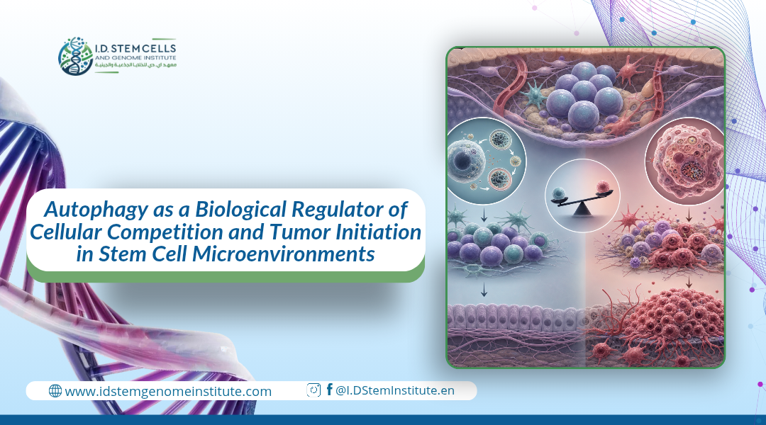
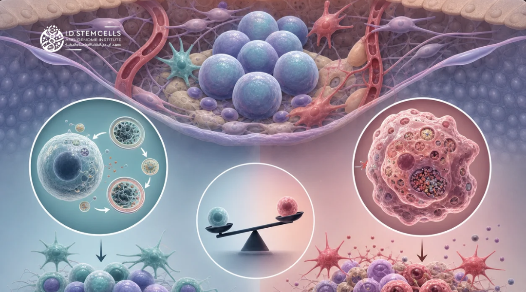
Adult stem cells undergo dynamic competition within their microenvironments (niches) to maintain tissue homeostasis. Cell displacement from these niches initiates differentiation. While healthy cells follow a neutral drift pattern through random cell replacement, cells carrying tumorigenic mutations gain a competitive advantage known as biased drift, enabling them to eliminate neighboring cells and initiate tumor formation. Autophagy is a vital metabolic mechanism for recycling cellular components. While its role in fueling tumor growth under stress conditions is well-established, its impact on stem cell spatial competition remains unclear. This study utilizes Drosophila ovarian germ cells as an ideal model due to their microstructure, which allows for monitoring competition at the single-cell level. Using this model, we aim to elucidate the role of autophagy in conferring competitive superiority to tumor-like cells within their niche.
Autophagy as a Key Driver of Tumor-like Stem Cell Dominance
1.Selective Competitive Advantage of BAM-Genetic Mutant Stem Cells
To assess the dynamics of competition between genetically distinct cell lineages within a niche, the FLP/FRT system was used to generate mosaic
Autophagy as a Key Driver of Tumor-like Stem Cell Dominance
1.Selective Competitive Advantage of BAM-Genetic Mutant Stem Cells
To assess the dynamics of competition between genetically distinct cell lineages within a niche, the FLP/FRT system was used to generate mosaic germ stem cells. The methodology relied on visual discrimination using green fluorescent protein (GFP), classifying microsites into three types: fully labeled (+GFP), mixed-labeled, and unlabeled (-GFP) sites dominated by mutant cells.
The study revealed that locus occupation was strongly skewed in favor of mutant stem cells in the bag of marbles (bam) gene; the percentage of sites colonized by these cells increased from 40% to 89% within 14 days. Interestingly, this competitive advantage was unique to bam cells and was not observed in other gene mutations such as brat, s, or stet. This functional difference is attributed to the biological nature of the cells; the bam mutation results in a homogeneous population of stem-like cells, while the other mutations lead to a mixture of stem and differentiated cells, confirming that locus dominance increases in homogeneous tumors.
2.Activation of the Autophagy Pathway in the Tumor Microenvironment
Given the known role of metabolic stress in stimulating cellular responses, autophagy activity in mutant cells was investigated using the fluorescent marker mCherry-Atg8a. The results showed significant variation in catabolic activity:
Control stem cells: exhibited very low levels of mCherry-Atg8a granules, reflecting minimal basal autophagy activity under normal conditions.
Baam gene mutant cells: showed a significant and pronounced increase in the number of fluorescent granules, indicating mobilization of the autophagy pathway.
3. Structural Confirmation via Transmission Electron Microscopy (TEM)
To reinforce these findings, high-resolution microscopic analyses were performed using transmission electron microscopy (TEM). The microscopic images confirmed that bam-mutant stem cells contain a large number of autophagic structures compared to control cells. This morphological and fluorescent evidence supports the hypothesis that tumor-like cells reprogram their metabolic system through autophagy to secure the resources necessary to outcompete and displace healthy cells from their microenvironment.
Tumor-like cells (mutant for the bam gene) exploit environmental stress conditions to their advantage. When fruit flies are exposed to nutrient deficiencies or "starvation," autophagy activity is significantly increased in these cells compared to healthy cells, as evidenced by the increased density of mCherry-Atg8a marker protein clusters. This metabolic stimulation induced by starvation was not merely a secondary response; it granted bam cells an additional competitive advantage, enabling them to occupy the specialized niche and displace normal cells more rapidly. To demonstrate that autophagy is the actual driver of this dominance, the researchers disabled the atg6 gene (responsible for this process) within the mutant cells, causing them to immediately lose their competitive advantage even under continued starvation conditions.
germ stem cells. The methodology relied on visual discrimination using green fluorescent protein (GFP), classifying microsites into three types: fully labeled (+GFP), mixed-labeled, and unlabeled (-GFP) sites dominated by mutant cells.
The study revealed that locus occupation was strongly skewed in favor of mutant stem cells in the bag of marbles (bam) gene; the percentage of sites colonized by these cells increased from 40% to 89% within 14 days. Interestingly, this competitive advantage was unique to bam cells and was not observed in other gene mutations such as brat, s, or stet. This functional difference is attributed to the biological nature of the cells; the bam mutation results in a homogeneous population of stem-like cells, while the other mutations lead to a mixture of stem and differentiated cells, confirming that locus dominance increases in homogeneous tumors.
2.Activation of the Autophagy Pathway in the Tumor Microenvironment
Given the known role of metabolic stress in stimulating cellular responses, autophagy activity in mutant cells was investigated using the fluorescent marker mCherry-Atg8a. The results showed significant variation in catabolic activity:
Control stem cells: exhibited very low levels of mCherry-Atg8a granules, reflecting minimal basal autophagy activity under normal conditions.
Baam gene mutant cells: showed a significant and pronounced increase in the number of fluorescent granules, indicating mobilization of the autophagy pathway.
3. Structural Confirmation via Transmission Electron Microscopy (TEM)
To reinforce these findings, high-resolution microscopic analyses were performed using transmission electron microscopy (TEM). The microscopic images confirmed that bam-mutant stem cells contain a large number of autophagic structures compared to control cells. This morphological and fluorescent evidence supports the hypothesis that tumor-like cells reprogram their metabolic system through autophagy to secure the resources necessary to outcompete and displace healthy cells from their microenvironment.
Tumor-like cells (mutant for the bam gene) exploit environmental stress conditions to their advantage. When fruit flies are exposed to nutrient deficiencies or "starvation," autophagy activity is significantly increased in these cells compared to healthy cells, as evidenced by the increased density of mCherry-Atg8a marker protein clusters. This metabolic stimulation induced by starvation was not merely a secondary response; it granted bam cells an additional competitive advantage, enabling them to occupy the specialized niche and displace normal cells more rapidly. To demonstrate that autophagy is the actual driver of this dominance, the researchers disabled the atg6 gene (responsible for this process) within the mutant cells, causing them to immediately lose their competitive advantage even under continued starvation conditions.
The Effect of Autophagy Deficiency on the Biological Characteristics and Lifecycle of Mutant Stem Cells
To elucidate the mechanism by which autophagy is impaired reduces the ability of mutant bam cells to occupy specialized sites, several potential physiological pathways were investigated. The results ruled out the linkage to adhesion proteins; no significant difference in E-cadherin protein levels was observed between normal cells, single-mutant bam cells, or double-mutant atg bam cells, thus negating the role of cell adhesion as the driver of this regression. Similarly, BMP signaling (measured via pMad levels) showed no difference to explain this competitive behavior, suggesting that autophagy-dependent competition operates via pathways independent of traditional growth signaling. Furthermore, the study demonstrated that autophagy impairment does not alter the "stem identity" of these cells; double-mutant cells continued to possess round spectrosomes, a hallmark of stem cells, without transforming into the branched spindle bodies associated with differentiation. Surprisingly, and contrary to the traditional role of autophagy in promoting survival, the TUNEL assay showed no increase in germ cell death rates even after 14 days of observation. Taken together, these data lead to the conclusion that autophagy in tumor-like cells does not function as a survival or adhesion mechanism, but rather as a biological regulator of cell cycle rate, providing the momentum necessary for numerical and spatial dominance within the microenvironment.
The Effect of Autophagy Deficiency on the Biological Characteristics and Lifecycle of Mutant Stem Cells
To elucidate the mechanism by which autophagy is impaired reduces the ability of mutant bam cells to occupy specialized sites, several potential physiological pathways were investigated. The results ruled out the linkage to adhesion proteins; no significant difference in E-cadherin protein levels was observed between normal cells, single-mutant bam cells, or double-mutant atg bam cells, thus negating the role of cell adhesion as the driver of this regression. Similarly, BMP signaling (measured via pMad levels) showed no difference to explain this competitive behavior, suggesting that autophagy-dependent competition operates via pathways independent of traditional growth signaling. Furthermore, the study demonstrated that autophagy impairment does not alter the "stem identity" of these cells; double-mutant cells continued to possess round spectrosomes, a hallmark of stem cells, without transforming into the branched spindle bodies associated with differentiation. Surprisingly, and contrary to the traditional role of autophagy in promoting survival, the TUNEL assay showed no increase in germ cell death rates even after 14 days of observation. Taken together, these data lead to the conclusion that autophagy in tumor-like cells does not function as a survival or adhesion mechanism, but rather as a biological regulator of cell cycle rate, providing the momentum necessary for numerical and spatial dominance within the microenvironment.
Autophagy as a Foundation for Competitive Superiority in Tumor Stem Cells
Our results confirm that autophagy plays a selective and crucial role in enabling tumor-like stem cells (bam gene mutant) to dominate their niche. While this catabolic pathway remains inactive and unnecessary for normal stem cells, it becomes vital for mutant cells to ensure their continued division and growth, reflecting a metabolic dependence similar to that observed in Ras gene-mutant cells.
Mechanism of Dominance: Division Rate, Not Adhesion Strength
Contrary to previous models that focused on the adhesion protein E-cadherin as a key factor in competition, our data indicate that the true advantage of bam cells lies in their cell cycle efficiency. Autophagy acts as a source of energy balance, allowing tumor-like cells to divide at rates exceeding those of their normal neighbors. It is noteworthy that this competition is directly influenced by tissue growth regulators such as insulin receptors and CDK2 kinase, making cancer stem cells more sensitive to external stimuli than epithelial cells.
Conclusion and Therapeutic Prospects: The study demonstrates that stem cell competition differs fundamentally from epithelial cell competition; the former relies on spatially displacing the opponent rather than killing it. Since autophagy promotes tumor growth in germ cells and supports their dominance over specialized sites, targeting this pathway represents a promising therapeutic strategy for disrupting tumor development in its early stages by depriving cancer cells of their competitive advantage.
At the I.D. Stem Cells and Genome Institute, we keep abreast of the latest scientific developments in the field of stem cell and gene therapy and regenerative medicine. If you are interested in learning more about the potential benefits of these treatments and the latest research findings, please feel free to contact us.
Autophagy as a Foundation for Competitive Superiority in Tumor Stem Cells
Our results confirm that autophagy plays a selective and crucial role in enabling tumor-like stem cells (bam gene mutant) to dominate their niche. While this catabolic pathway remains inactive and unnecessary for normal stem cells, it becomes vital for mutant cells to ensure their continued division and growth, reflecting a metabolic dependence similar to that observed in Ras gene-mutant cells.
Mechanism of Dominance: Division Rate, Not Adhesion Strength
Contrary to previous models that focused on the adhesion protein E-cadherin as a key factor in competition, our data indicate that the true advantage of bam cells lies in their cell cycle efficiency. Autophagy acts as a source of energy balance, allowing tumor-like cells to divide at rates exceeding those of their normal neighbors. It is noteworthy that this competition is directly influenced by tissue growth regulators such as insulin receptors and CDK2 kinase, making cancer stem cells more sensitive to external stimuli than epithelial cells.
Conclusion and Therapeutic Prospects: The study demonstrates that stem cell competition differs fundamentally from epithelial cell competition; the former relies on spatially displacing the opponent rather than killing it. Since autophagy promotes tumor growth in germ cells and supports their dominance over specialized sites, targeting this pathway represents a promising therapeutic strategy for disrupting tumor development in its early stages by depriving cancer cells of their competitive advantage.
At the I.D. Stem Cells and Genome Institute, we keep abreast of the latest scientific developments in the field of stem cell and gene therapy and regenerative medicine. If you are interested in learning more about the potential benefits of these treatments and the latest research findings, please feel free to contact us.