.png)
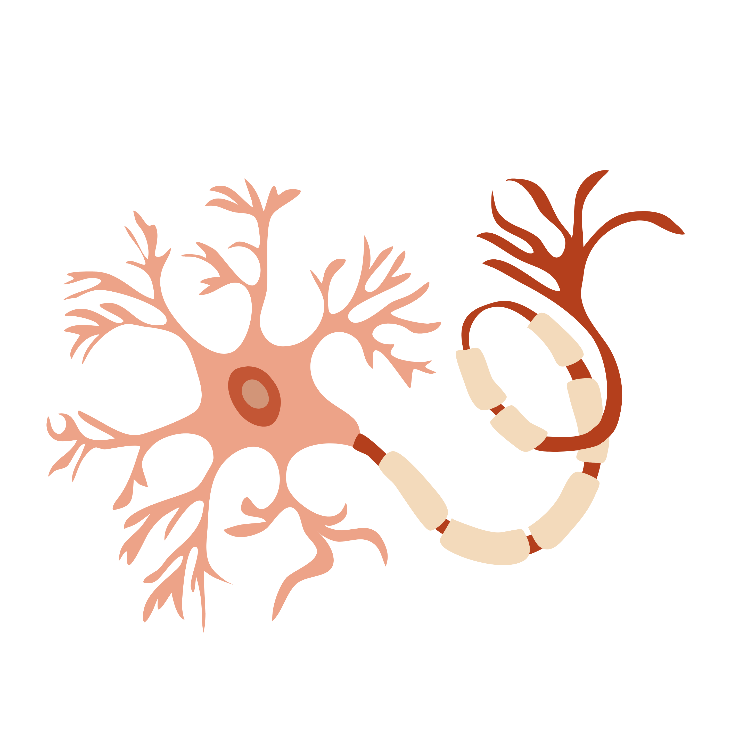
The development of 3D models of neurons began in the 1990s when neurospheres were formed using neural progenitor cells.
Neurospheres are 3D structures containing neurons and glial cells but lacking the organized cellular architecture of brain tissue. A major breakthrough in the development of 3D models occurred a few years after the advent of iPSCs when Lancaster and colleagues developed the first successful brain organoid protocol. Numerous researchers worldwide have refined and improved 3D culture protocols directed to the in vitro formation of specific cortical regions from different types of iPSCs. Brain organoids can be produced using both undirected and directed methods.
Undirected protocols rely on cell differentiation and self-organization with minimal induction factors, resulting in the formation of brain organoids, which are composed of different brain regions and exhibit significant heterogeneity between organoids and batches. In targeted approaches, induction factors are used to direct differentiation toward specific brain regions of interest or regional neural organoids.
Microfibrils at the embryoid body (EB) stage can enhance neuroectoderm formation and promote cortical development. The cortical region of these organoids offers better formation of the polarized cortical plate and radial units compared to undirected brain organoids.
Using induction factors, it is also possible to generate forebrain organoids, including the subplate, cortical plate, and Cajal-Retzius cell zones—in addition to the three progenitor zones—the ventricular, subventricular, and intermediate zones. After minor modifications, this model has recently been used to analyze the impact of genetic mutations on neural development.