Medical Articles

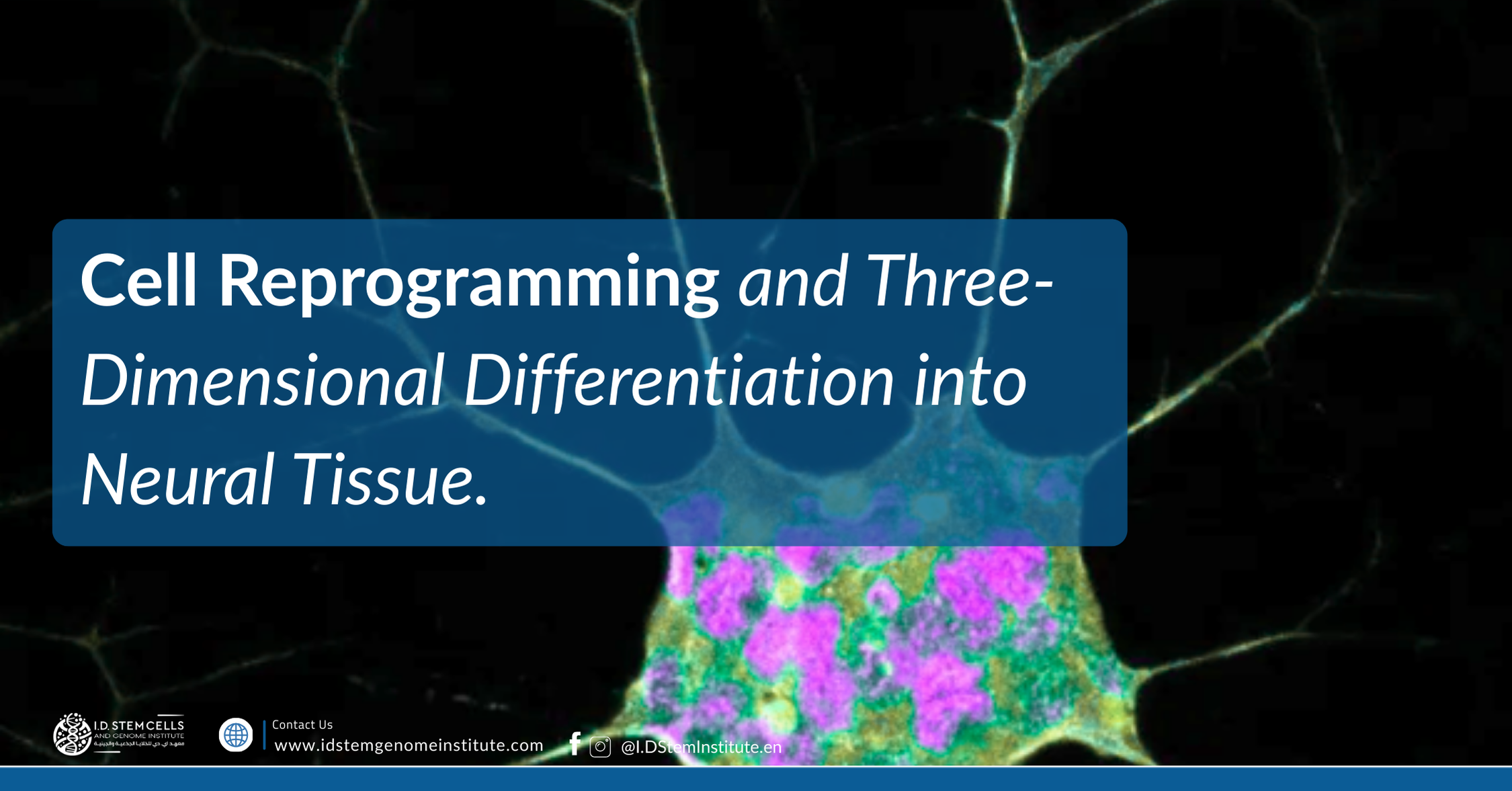
Cell Reprogramming and Three-Dimensional Differentiation into Neural Tissue

Cell Reprogramming and Three-Dimensional Differentiation into Neural Tissue

The use of patient-derived stem cells to model neurodevelopmental diseases is not new, having begun with the advent of somatic cell reprogramming technology.

iPSCs retain the genetic background of their donors and have been widely applied in studies of ASD through two-dimensional models of differentiation into neural progenitor cells (NPCs), neurons, astrocytes, and microglia. These studies have compared the phenotypes of differentiated cells between patients with ASD and controls, which can be unrelated or related to the neurological subjects, or even CRISPR-edited isogenic controls, in which the mutation of interest is introduced or corrected. Recently, brain organoids have emerged as an important model for analyzing many aspects of the neurobiology
Image

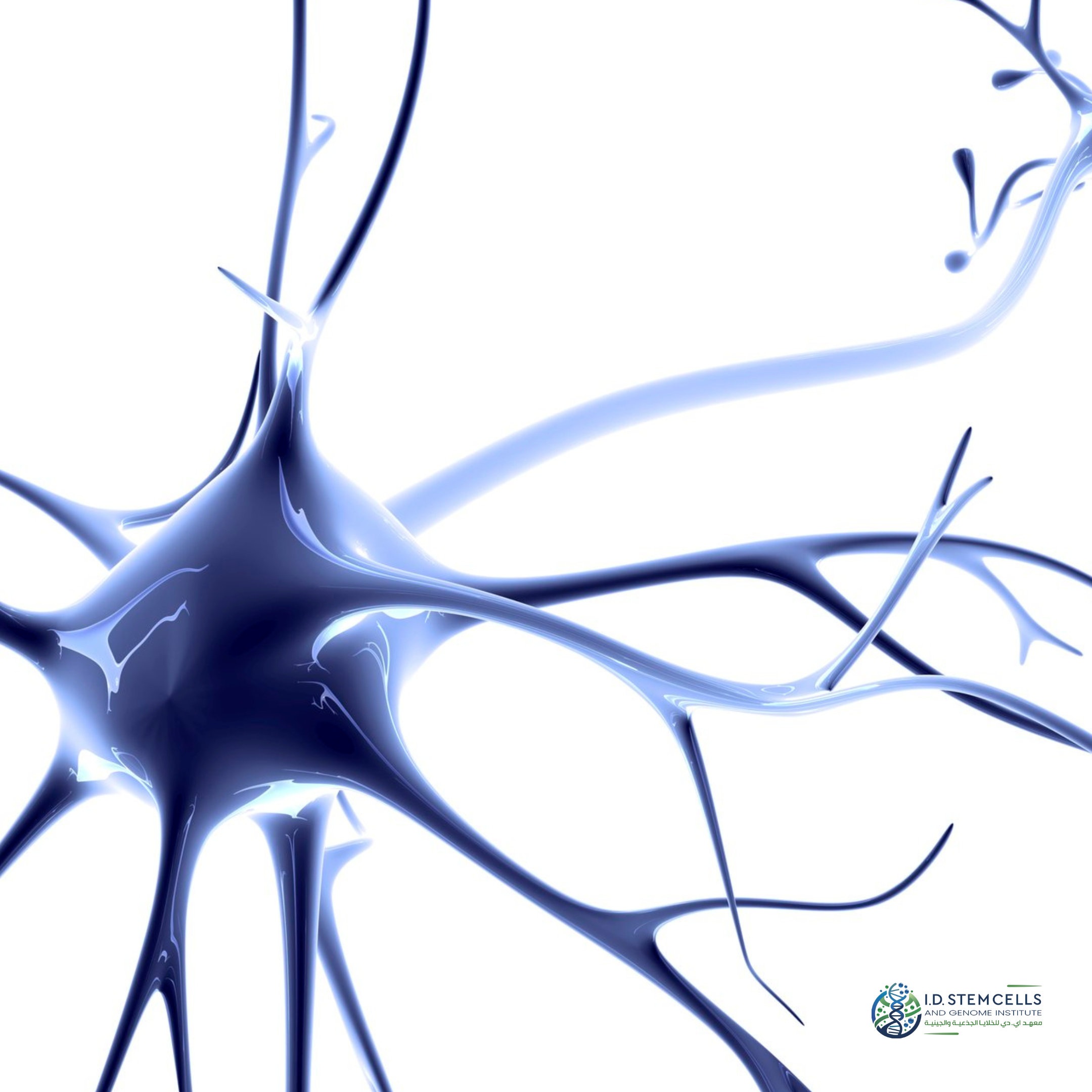
of this neuropsychiatric disorder because they offer improved in vitro reproducibility to capture the complexity of the events and cellular interactions present in early neurodevelopment. Cell reprogramming and three-dimensional differentiation into neural tissue. Somatic cells from patients, such as fibroblasts and peripheral blood mononuclear cells (PBMC), undergo cellular reprogramming through the introduction of Yamanaka factors. These cells become induced pluripotent stem cells (iPSCs) and are used to create three-dimensional structures such as brain organoids and spheroids.

Brain organoids provide a cellular architecture that better recapitulates what is observed during organogenesis in vivo. Therefore, brain organoids are excellent tools for addressing some of the gaps left by two-dimensional models and better reproducing key features of the developing human brain. Interestingly, gene expression and co-expression network analyses have identified autism spectrum disorder risk genes in the development of excitatory and inhibitory neurons in the inner cortical plate, subventricular zone, and subiculum during mid-gestation and developmental stages, and biological processes that can be modeled using brain organoids


For immediate consultation with experts from the I.D. Institute for Stem Cell and Gene Research

For immediate consultation with experts from the I.D. Institute for Stem Cell and Gene Research

Image

you may also like

Image

you may also like

Image
Image
Image
Image

I.D. Journal of Stem Cell Research and Advanced Therapeutics

A medical journal published by the I.D. Institute for Stem Cell and Genome Research

Image
Image
Image
Image
Image
Image
Image
Image
I.D. Journal of Stem Cell Research and Advanced Therapeutics

A medical journal published by the I.D. Institute for Stem Cell and Genome Research

Image
Image
Image
Image
Image

I.D. Educational Community

Join the I.D. Community, an interactive environment bringing together experts, alumni, and students. This community aims to:

  • Exchange Clinical Expertise: Discuss challenging medical cases to enhance treatment outcomes.

  • Discuss Cutting-edge Research: Explore contemporary debates such as Digital Twins, Gerontology, and AI in medicine.

  • Build Professional Networks: Connect with leaders in regenerative medicine and global health organizations.

Image
Image
Image

I.D. Educational Community

Join the I.D. Community, an interactive environment bringing together experts, alumni, and students. This community aims to:

  • Exchange Clinical Expertise: Discuss challenging medical cases to enhance treatment outcomes.

  • Discuss Cutting-edge Research: Explore contemporary debates such as Digital Twins, Gerontology, and AI in medicine.

  • Build Professional Networks: Connect with leaders in regenerative medicine and global health organizations.

Image
Image
Image
Image

© 2025 I.D. Holding, By prof. Dr. Islam Dababseh

© 2025 I.D. Holding, By prof. Dr. Islam Dababseh

Image

© 2025 I.D. Holding, By prof. Dr. Islam Dababseh

© 2025 I.D. Holding, By prof. Dr. Islam Dababseh

Image