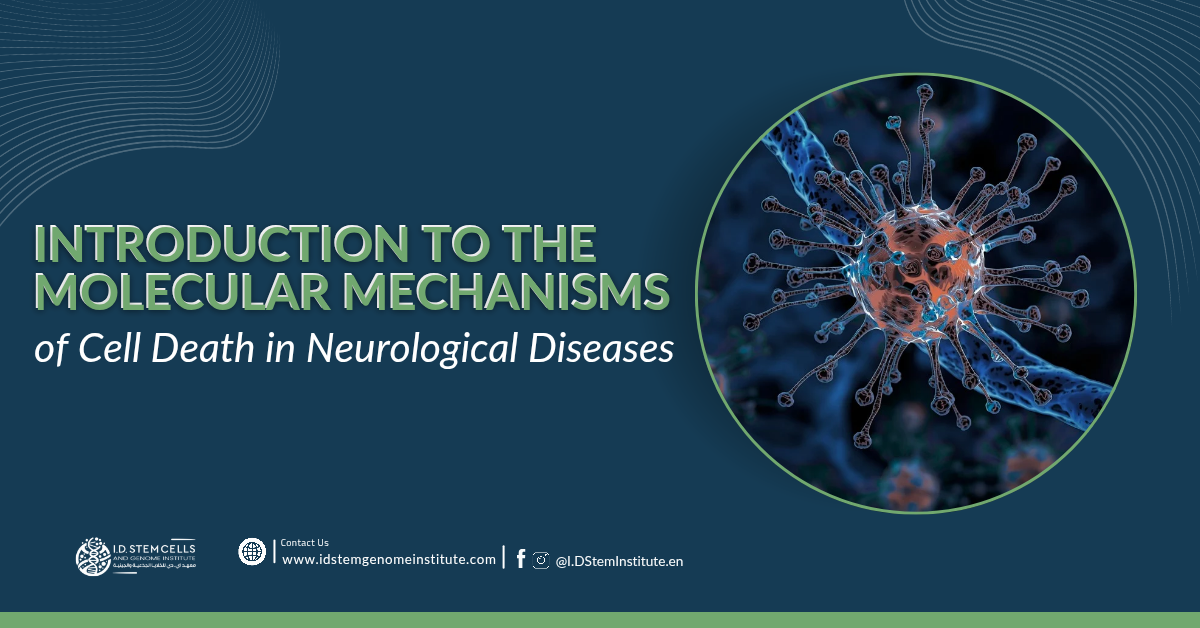
Programmed cell death (PCD) is required for normal development and maintenance of tissue homeostasis, eliminating damaged, injured, or aging cells in multicellular organisms.
In 1972, he identified the structurally distinctive features of cells undergoing programmed suicide, coining the term "apoptosis" for this type of PCD. These features include cytoplasmic shrinkage, nuclear condensation and fragmentation, and the formation of apoptotic bodies, which appear in various tissues under certain physiological or pathological conditions.
A family of anti- and pro-apoptotic BCL-2 (BCL-2) protein members was discovered that regulates this pathway. These members are divided into three subgroups based on their structure and function, with conserved regions called BCL-2 homology (BH) motifs. These include anti-apoptotic proteins (BCL-2, BCL-XL, MCL-1, BCL-W, and A1/BFL1), BH3-only proteins (BIM, PUMA, BID, BMF, BAD, HRK, BIK, and NOXA), critical initiators of apoptosis, and multi-BH domain proteins (BAX and BAK), essential effectors of apoptosis that form oligomers that cause mitochondrial outer membrane permeabilization (MOMP), thereby releasing apoptotic factors that promote a cascade of caspases (aspartate-activated cysteine-specific proteinases). Upon activation, caspases cleave hundreds of cellular substrates, precipitating the morphological hallmarks of apoptosis and cell destruction.
Normal development and tissue homeostasis in multicellular organisms depend on tightly regulated, coordinated PCD signaling events. During embryonic development, cell death by PCD is essential for the proper formation of certain tissues, for example, the sculpting of vertebrate limb digits. The central nervous system (CNS), consisting of the brain and spinal cord, is formed by PCD, where tightly regulated signaling events at both a temporal and spatial level lead to the establishment of neural architecture. In normal embryonic and postnatal neural development, apoptosis is the main form of PCD. Apoptosis can affect various cell populations, including neural progenitor cells (NPCs), post-mitotic neurons, and glial cells, ensuring that only cells of the correct size and shape survive, and that have established appropriate connections with their axons and neurons. In mouse embryos, neurogenesis occurs as early as E12 when NPCs exit the cell cycle and differentiate into post-mitotic neurons. Members of the anti-apoptotic BCL-2 family, myeloid leukemia-1 (MCL-1) and BCL-2-associated long motif (BCL-XL), have been shown to play critical roles in cell survival during developmental neurogenesis.